سانتریول
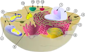
الگو:زیستشناسیی سلولی در زیست سلولی، سانتریول اندامک نیست و عمدتاً از پروتئینهایی به نام توبولین تشکیل شدهاست.[1] سانتریولها در بسیاری از سلولهای یوکاریوتی یافت میشوند. یک جفت سانتریول که در محاصره توده ماده چگالی به نام ماده پیرامون مرکز (PCM) قرار داشته باشد، تشکیل ساختاری به نام سانتروزوم را میدهد.[1]
سانتریولها در تمام سلولهای یوکاریوتی حضور ندارند، برای نمونه در مخروطیان، گیاهان گلدار و بسیاری از قارچها غایباند و تنها در گامتهای نر سنگخزهتباران (کاروفیتها)، خزهتباران (بریوفیتها)، گیاهان آونددار بدون دانه، سرخس نخلی و کهندارها حضور دارند.[2][3]
سانتریولها اغلب از نه دسته میکروتوبولهای سهگانه که به صورت استوانه ای آرایش یافتهاند تشکیل شدهاند. مواردی که از این ساختار انحراف پیدا کردهاند شامل این موارد میشود: رویان خرچنگها و مگس سرکه با نه جفت میکروتوبول، سلولهای اسپرم کرم الگانس و جنین اولیه آن با نه میکروتوبول تکین.[4][5]
عملکرد اصلی سانتریولها در تولید مژک طی اینترفاز و استر و دوک طی تقسیم سلولی میباشد.
تاریخچه
ادوارد ون بندن سنتروزوم (میانتن)ها را اولین بار در ۱۸۸۳ مشاهده کرد.[6] در ۱۸۹۵، تئودور بووری این اندامک را «سنتروزوم» نامید.[7][8] الگوی تکثیر سانتریول اولین بار توسط اتین دو هارون و جوزف جی. گال در ۱۹۵۰ بدست آمد.[9][10]
منابع
- Eddé, B; Rossier, J; Le Caer, JP; Desbruyères, E; Gros, F; Denoulet, P (1990). "Posttranslational glutamylation of alpha-tubulin". Science. 247 (4938): 83–5. Bibcode:1990Sci...247...83E. doi:10.1126/science.1967194. PMID 1967194.
- Quarmby, LM; Parker, JD (2005). "Cilia and the cell cycle?". The Journal of Cell Biology. 169 (5): 707–10. doi:10.1083/jcb.200503053. PMC 2171619. PMID 15928206.
- Silflow, CD; Lefebvre, PA (2001). "Assembly and motility of eukaryotic cilia and flagella. Lessons from Chlamydomonas reinhardtii". Plant Physiology. 127 (4): 1500–1507. doi:10.1104/pp.010807. PMC 1540183. PMID 11743094.
- Delattre, M; Gönczy, P (2004). "The arithmetic of centrosome biogenesis" (PDF). Journal of Cell Science. 117 (Pt 9): 1619–30. doi:10.1242/jcs.01128. PMID 15075224.
- Leidel, S.; Delattre, M.; Cerutti, L.; Baumer, K.; Gönczy, P (2005). "SAS-6 defines a protein family required for centrosome duplication in C. elegans and in human cells". Nature Cell Biology. 7 (2): 115–25. doi:10.1038/ncb1220. PMID 15665853.
- Wunderlich, V. (2002). "JMM - Past and Present". Journal of Molecular Medicine. 80 (9): 545–548. doi:10.1007/s00109-002-0374-y. PMID 12226736.
- Boveri, T. Ueber das Verhalten der Centrosomen bei der Befruchtung des Seeigel-Eies nebst allgemeinen Bemerkungen über Centrosomen und Verwandtes. Verh. d. Phys. -Med. Ges. zu Würzburg, N. F. , Bd. XXIX, 1895. link.
- Boveri, T. (1901). Zellen-Studien: Uber die Natur der Centrosomen. IV. Fischer, Jena. link.
- Wolfe, Stephen L. (1977). Biology: the foundations (First ed.). Wadsworth. ISBN 978-0-534-00490-3.
- Vorobjev, I. A.; Nadezhdina, E. S. (1987). The Centrosome and Its Role in the Organization of Microtubules. International Review of Cytology. 106. pp. 227–293. doi:10.1016/S0074-7696(08)61714-3. ISBN 978-0-12-364506-7. PMID 3294718.. See also de Harven's own recollections of this work: de Harven, Etienne (1994). "Early observations of centrioles and mitotic spindle fibers by transmission electron microscopy". Biol Cell. 80 (2–3): 107–109. doi:10.1111/j.1768-322X.1994.tb00916.x. PMID 8087058.